X-ray of Front Teeth Implants: Front teeth inserts are now a more common option for each looking for a lasting fix for front teeth that are missing or damaged. Dental inserts provide a long-lasting solution that can bring back both the look and function naturally. Yet, for the procedure to be successful, dental masters depend on advanced imaging methods, such as X-rays, to thoroughly judge and oversee the process.

This article examines the importance of X-rays in front teeth inserts, how the process works, and what patients can expect throughout the journey. By understanding these explanations, patients can feel more ready and confident about their decisions.
What is an X-ray in Dental inserts?
X-rays play a difficult role in planning, placing, and maintaining dental inserts, especially for front teeth. An X-ray is a diagnostic scanning tool that uses controlled radiation to capture deep images of bone, teeth, and other systems within the mouth. For dental inserts, X-rays allow dentists to test the jawbone and the surrounding areas, helping to secure a stable foundation for the insert.
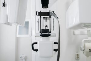
Various kinds of X-rays, such as broad, periapical, and CBCT scans, are employed in dental insert procedures. Each kind provides unique advantages and helps dentists in developing a more precise treatment process.
The Importance of X-Rays for Dental Implants in Front Teeth
- Front teeth implants require X-rays as they reveal important information that is not visible through a regular dental check-up. This is why X-rays are necessary.
- Assessment of the jawbone using X-rays is crucial for dentists to evaluate its density and volume to ensure a secure anchoring of the implant.
- Precise placement of the implant is important for front teeth because they are very visible. X-rays assist in determining the perfect positioning.
- Discovery of Nerve Pathways: Utilizing X-rays to pinpoint nerve positions can help avoid complications when inserting implants.
- Dentists use X-rays to check for any hidden disease or decline that could impact the success of the implant.
| X-Ray Type | Purpose | Details |
|---|---|---|
| Panoramic X-Ray | Initial assessment | Provides a broad view of the jawbone and teeth |
| Periapical X-Ray | Focused monitoring | Detailed view of specific teeth and bone structure |
| CBCT Scan | 3D imaging and precision planning | Offers highly detailed 3D images for accurate implant placement |
Different kinds of X-rays are utilized for front teeth implants.
Various X-ray techniques can be used while carrying out the implant procedure. Every kind has a unique function, offering dentists the additional information required during every phase of the implant process.
1. Broad X-Rays
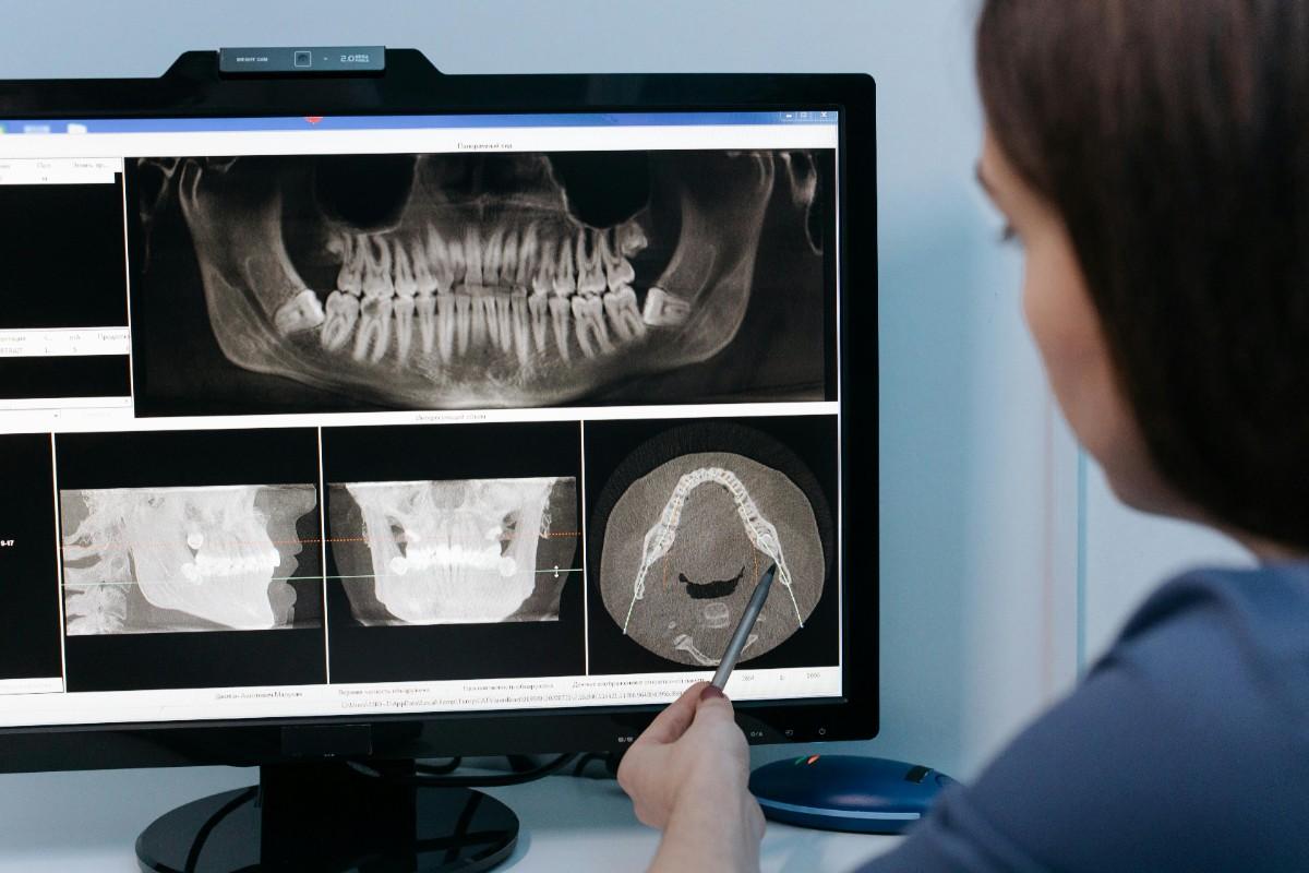
Broad X-rays capture a wide view of the entire mouth, including the jawbone, sinuses, and teeth. This broad view is useful for calculating the general bone system and finding any underlying issues. Broad X-rays are commonly taken at the initial meeting to help plan the treatment.
2. Apical X-Rays
Apical X-rays focus on a specific area, usually one or two teeth at a time. These X-rays provide a detailed view of the tooth and surrounding bone structure, making them helpful for observing insert progress. Apical X-rays are often used during follow-up visits after the insert is placed.
3. Cone Beam CT (CBCT) Reviews
CBCT review offers 3D scanning, giving dentists a highly detailed view of the jawbone, nerves, and surrounding system. For front teeth inserts, CBCT scans are critical because they allow precise measurements and reveal any potential obstacles in the bone. This advanced imaging technique is often used for complex cases.
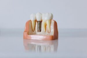
The Implant Process and Role of X-Rays at Each Step
Placing front teeth implants involves several steps, and dentists use X-rays throughout to ensure each stage is completed successfully. Here’s an overview of how X-rays play a role in each phase:
1. Initial Consultation and Planning
During the first consultation, dentists take panoramic or CBCT X-rays to evaluate overall bone health and design. This step helps the dentist see whether a bone graft is vital and plan the precise location for the insert order.
2. Bone Joining, If Required
If the first X-rays show that the jawbone is without thickness, the dentist may recommend a bone transplant. In such cases, dentists take additional X-rays after the grafting process to watch bone growth and ensure the jaw is ready for implant arrangement.
3. Insert Placement Surgery
On the day of surgery, dentists use X-rays to locate and confirm the exact spot. Once the implants are set in the bone, they may take additional X-rays to ensure that everything is securely in place.
4. Healing and Osseointegration
After the implants are placed, the bone needs time to fuse with the implant—a process known as osseointegration. Dentists often take X-rays during follow-ups to monitor progress and confirm that the implant is integrating successfully with the bone.
5. Attaching the Crown
Once the implant has integrated with the bone, the dentist attaches a crown to the implant post. Before attaching the crown, they take final X-rays to ensure proper alignment and fit.
Advantages of Using X-Rays for Front Teeth Implants
The use of X-rays in front teeth implants provides numerous benefits, both for the dentist and the patient:
- Improved Accuracy: X-rays provide detailed images that help dentists place inserts with identified precision, necessary for front teeth.
- Reduced Risk of Complications: By determining nerve pathways and bone density, X-rays help protect against issues like nerve damage or insert failure.
- Enhanced Safety: With detailed scanning, dentists can make better decisions, minimizing the likelihood of post-surgical complications.
- Better Aesthetics: Detailed insert placement is key for front teeth, where look matters. X-rays help verify the implant aligns well with natural teeth for a smooth look.
- Risks and Safety Measures of X-Rays in Dental Implants While X-rays use controlled radiation, they are normally safe for dental procedures. Dentists take measures to reduce uncovering, such as using lead overall and limiting the number of X-rays taken. CBCT scans, though powerful, are only used when essential to reduce radiation uncovering.

Patients should always notify their dentist if they are pregnant or have worries about radiation. In most cases, the advantages of X-rays for insert planning far prevail over the risks, as they help verify a safe, successful result.
What Patients Can Assume During an X-Ray for Front Teeth Implants?
The X-ray procedure is fast and usually painless for the majority of patients. During the process:
Placing: You’ll be asked to sit or stand while the X-ray machine is adjusted to catch the best angle.
Radiation Protection: The dentist may cover you with a lead apron to protect other areas of your body from uncovering.
Fast Scanning: The current X-ray scan is completed within a matter of seconds.
Following that, the dentist will go over the images and have a talk with you about them, helping you grasp the upcoming stages of the implant procedure.
How to Prepare for an X-Ray and Implant Procedure
There isn’t much preparation needed for an X-ray, but for the insert surgery, patients need to fulfill some tasks:
Notify the Dentist: Make sure your dentist is updated on any medical conditions, sensitivities, or treatment.
Shun Eating or Drinking: Depending on the pain relief used, your dentist may advise you to avoid eating or drinking a few hours before surgery.
Arrange Transportation: For the insert process, arrange for someone to drive you home if calming is used.
Common Questions About X-Rays and Front Teeth Implants
1. Are X-rays necessary for every insert?
Yes, X-rays are essential for insert success, as they help evaluate the bone system and monitor progress.
2. Will insurance cover X-rays for dental inserts?
Many insurance plans cover X-rays, but it’s best to check with your provider as the scope changes.
3. How often will I need X-rays during the insert process?
Dentists usually take X-rays during the first consultation, after bone grafting (if necessary), on the day of implant surgery, and during follow-up appointments.
Summary: The Importance of X-Rays in Confirm Successful Insertion of Front Teeth
- X-rays are important for the location of front teeth as they give dentists an exact view of the jaw and surrounding areas. Dentists use their experience to apply X-rays, ensuring that every step, from the initial planning to the placement of the final crown, is completed with great attention to detail. This will guide to a better experience for patients, as well as an enhanced length of life of insert and a beautiful, genuine smile that gains confidence and quality of life.
- A front teeth insert can transform a person’s look and oral health. With the help of X-rays, patients can gain the best possible result, supported by cutting-edge technology and master care.

1 thought on “X-Ray of Dental Insert in Front Teeth: Overview of the Process and its Significance.”